What is Dental Cone Beam CT?
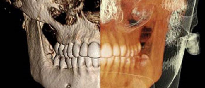
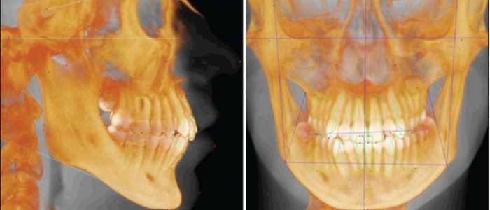
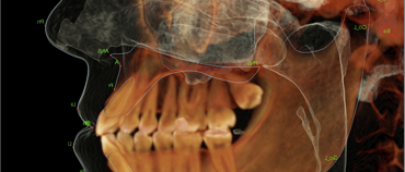
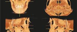
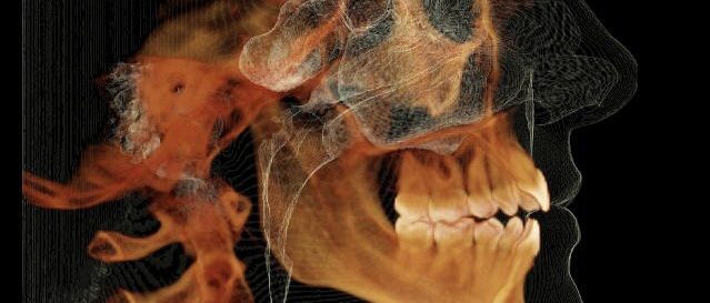
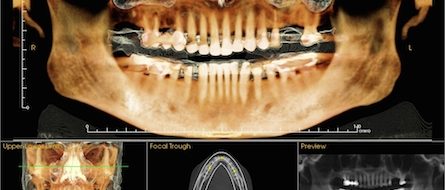
Dental cone beam computed tomography (CT) is a special type of x-ray machine used in situations where regular dental or facial x-rays are not sufficient. It is not used routinely because the radiation exposure from this scanner is significantly more than regular dental x-rays. See the Safety page for more information about x-rays. This type of CT scanner uses a special type of technology to generate three dimensional (3-D) images of dental structures, soft tissues, nerve paths and bone in the craniofacial region in a single scan. Images obtained with cone beam CT allow for more precise treatment planning.

You should wear comfortable, loose-fitting clothing to your exam. You may be given a gown to wear during the procedure.
Metal objects, including jewelry, eyeglasses, dentures and hairpins, may affect the CT images and should be left at home or removed prior to your exam. You may also be asked to remove hearing aids and removable dental work. Women will be asked to remove bras containing metal underwire. You may be asked to remove any piercings, if possible.
Cone beam CT is not the same as conventional CT. However, dental cone beam CT can be used to produce images that are similar to those produced by conventional CT imaging.
With cone beam CT, an x-ray beam in the shape of a cone is moved around the patient to produce a large number of images, also called views. CT scans and cone beam CT both produce high-quality images.
Dental cone beam CT was developed as a means of producing similar types of images but with a much smaller and less expensive machine that could be placed in the dentist’s office.
Cone beam CT provides detailed images of the bone and is performed to evaluate diseases of the jaw, dentition, bony structures of the face, nasal cavity and sinuses. It does not provide the full diagnostic information available with conventional CT, particularly in evaluation of soft tissue structures such as muscles, lymph nodes, glands and nerves. However, cone beam CT has the advantage of lower radiation exposure compared to conventional CT.
What are some common uses of the procedure?
Dental cone beam CT is commonly used for treatment planning of orthodontic issues. It is also useful for more complex cases that involve:
surgical planning for impacted teeth.
diagnosing temporomandibular joint disorder (TMJ).
accurate placement of dental implants.
evaluation of the jaw, sinuses, nerve canals and nasal cavity.
detecting, measuring and treating jaw tumors.
determining bone structure and tooth orientation.
locating the origin of pain or pathology.
cephalometric analysis.
reconstructive surgery.
What are the Benefits vs Risks
Benefits
- The focused x-ray beam reduces scatter radiation, resulting in better image quality.
- A single scan produces a wide variety of views and angles that can be manipulated to provide a more complete evaluation.
- Cone beam CT scans provide more information that conventional dental x-ray, allowing for more precise treatment planning.
- CT scanning is painless, noninvasive and accurate.
- A major advantage of CT is its ability to image bone and soft tissue at the same time.
- No radiation remains in a patient’s body after a CT examination.
- X-rays used in CT scans should have no immediate side effects.
Risks
- There is always a slight chance of cancer from excessive exposure to radiation. However, the benefit of an accurate diagnosis far outweighs the risk.
- CT scanning is, in general, not recommended for pregnant women unless medically necessary because of potential risk to the baby in the womb.
- Because children are more sensitive to radiation, they should have a CT exam only if it is essential for making a diagnosis and should not have repeated CT exams unless absolutely necessary. CT scans in children should always be done with low-dose technique.